Medical Terminology Daily (MTD) is a blog sponsored by Clinical Anatomy Associates, Inc. as a service to the medical community. We post anatomical, medical or surgical terms, their meaning and usage, as well as biographical notes on anatomists, surgeons, and researchers through the ages. Be warned that some of the images used depict human anatomical specimens.

You are welcome to submit questions and suggestions using our "Contact Us" form. The information on this blog follows the terms on our "Privacy and Security Statement" and cannot be construed as medical guidance or instructions for treatment.


We have 314 guests online


A Moment in History

Jean-Louis Petit

Jean Louis Petit
(1674 – 1750)

French surgeon and anatomist, Jean Louis Petit was born in Paris in on March 13, 1674.  His family rented an apartment at his house to Alexis Littre (1658 – 1726), a French anatomist. Petit became an apprentice of Littre at seven years of age, helping him in the dissections for his lectures and at an early age became the assistant in charge of the anatomic amphitheater.

Because of Petit’s dedication to anatomy and medicine, in 1690 at the age of sixteen, became a disciple of a famous Paris surgeon, Castel.

In 1692, Petit entered the French army and performed surgery in two military campaigns. By 1693 he started delivering lectures and was accepted as a great surgeon, being invited to the most difficult operations.  In 1700 he was appointed Chief Surgeon of the Military School in Paris and in the same year he received the degree of Master of Surgery from the Faculty of Paris.

In 1715 he was made a member of the Royal Academy of Sciences and an honorary member of the Royal Society of London. He was appointed by the King as the first Director General of the Royal Academy of Surgery when it was founded in 1731.

Petit’s written works are of historical importance.  “Traite des Maladies des Os” ( A Treatise on Bone Diseases);  “Traite des Maladies Chirurgicales et des Operation” (A Treatise on Surgical Diseases and their Operations” This last book was published posthumously in 1774. He also published a monograph on hemorrhage, another on lachrymal fistula, and others.

He was one of the first to perform choIecystotomy and mastoidotomy. His original tourniquet design for amputations saved many in the battlefield and the design of the same surgical instrument today has not changed much since its invention by him.

His name is remembered in the lumbar triangle, also called the "triangle of Petit", and the abdominal hernia that can ensue through that area of weakness, the lumbar hernia or "Petit's hernia".

Sources:
1. “Jean Louis Petit – A Sketch of his Life, Character, and Writings” Hayne, AP San Fran Western Lancet 1875 4: 446-454
2. “Oeuvres compl?tes de Jean-Louis Petit” 1837 Imprimerie de F. Chapoulaud
3. Extraits de l'eloge de Jean-Louis Petit Ius dans Ia seance publique de I' Academie royale de chirurgie du 26 mai 1750” Louis A. Chirurgie 2001: 126 : 475- 81


 "Clinical Anatomy Associates, Inc., and the contributors of "Medical Terminology Daily" wish to thank all individuals who donate their bodies and tissues for the advancement of education and research”.

Click here for more information


abebooks banner

bookplateink.com


The Azygos Lobe


This article was written by Claudio Rodrigo Molina, Josefa Catalán Lobo, and Carolina Becker Fehlandt. We thank them for their contribution to Medical Terminology Daily.


The azygos lobe, also commonly referred to as the" accessory lobe of the azygos vein", was first described in 1877 by Heinrich Wrisberg. It is seen in 0,4 % of chest X-rays; 1,5% in high resolution CT, and 1% in anatomical dissections. It is not a true accessory pulmonary lobe as it does not have its own bronchus and does not correspond to a specific bronchopulmonary segment, the “azygos lobe”, is located at the apicomedial portion of the upper right lobe and it separated from the remainder of the upper lobe by a fissure (Denega et al, 2015; Özdemir et al, 2017)

A convex-shaped fissure is created by the course of the vein bearing towards the medial side of the right lung to join with the superior vena cava. Its formation is a result of an unusual embryogenic migration of the posterior cardinal vein; which is a precursor of the azygos vein (Denega et al, 2015).

Instead of sliding over the lung medially, the vein invaginates into the parenchyma of the lung and becomes enveloped by layer of pleural folds, forming a mesentery-like structure, also called “mesoazygos”. Further migration into the lung as it passes towards the right hilum creates a convex semicircular fissure with the vein located at the base of the fissure. This fissure can be identified in an X-ray chest image as a coma-shaped (“Teardrop sign”) or curved linear shadow in the paramediastinal region of the right lung; it terminates at the level of second costal cartilage (Akhtar et al, 2018; Caceres et al, 1993). The lower portion of the azygos fissure is teardrop-shaped and its contains the azygos vein (Özdemir et al, 2017; Kotovet al, 2017).

The pathway of the vein within the lung is subject to individual variation, and this defines the position of the fissure within the apex of the upper lobe. The most superior portion of the fissure adopts a triangular form, called the trigone. The localization of the trigone determinates the size of the azygos lobe (Caceres et al, 1993; Fuad & Mubarak, 2016 ). A Left azygos lobe has been reported, but it is extremely rare (Özdemir et al, 2017)

Diagnosis of the azygos lobe may be complicated by morphologic variants of the fissure, physiological changes in the size of vein, and the projection of additional shadows within the lobe which may be misinterpreted as scar tissue, a calcified area of a post-infection process, or a malignant tissue or nodule.

 Lateral view, the arrow shows the location of the azygos lobe

Anterior view, the arrow shows the location of the azygos lobe
Images provided by the authors.  Click on the image for a larger depiction

It usually has no clinical implications and is an incidental finding in images but the azygos vein may undergo physiological variations, reflected by changes in the size of its shadow and its position in the imaging studies. Expiration, the Valsalva maneuver, or the upright position effect on the venous return to the heart, may enhance the size of the vein and its shadow. Changes in intrathoracic pressure may result in the “empty azygos fissure” phenomenon, in which the medial displacement of the azygos vein occurs after the reexpansion of the collapsed lung, secondary to pneumothorax or pleural effusion as well as a shortened mesoazygos.

In rare cases the azygos veins may undergo variceal changes that are usually located in the arc of the vein. They remained asymptomatic or may be accompanied by a “pressure like” or “tightness” sensation within the chest, recurrent hemoptysis with bright red blood dry cough and dyspnea. The initial differential diagnosis includes acute myocardial infarction, aortic dissection and myocarditis. On the chest X-ray it may present as a round or oval paratracheal shadow with a smooth surface or outline. Untreated, it may predispose the patient to the risk of rupture, thrombosis, or pulmonary embolism. Azygos thrombosis is extremely rare and most cases in the literature had an undelaying azygos dilation or some prothrombotic status like malignancy. In all lung tissue some pathological process can originate in the azygos lobe as bullous, bronchiectatic changes, pneumonia and tuberculosis. (Kotov et al, 2018; Denega et al, 2015)

Otherwise, it seems the mesoazygos fold serve as a barrier to dissemination of the infection or malignant cells.

For thoracoscopic procedures, recognition of the azygos lobe is particularly important as it can cause partial obstruction of the surgical site view during thoracoscopic sympathectomy. In the literature, two cases have been reported where the phrenic nerve was coursing within the azygos fissure (Kauffman et al, 2010; Pradhan, 2017; Özdemir et al, 2017; Paul, Siba & James, 2018)

Thoracic surgeons as well as treating physicians need to be aware of this rare anatomical variation.

NOTE: For an explanation of the etymology of the word "azygos" click here.

Sources:

1. Akhtar, Jamal & Lal, Amos & B. Martin, Kevin & Popkin, Joel. (2018). Azygos lobe: A rare cause of right paratracheal opacity. Respiratory Medicine Case Reports. 23. 10.1016/j.rmcr.2018.02.001.
2. Caceres, Jennelyn & Mata, Jonathan & Alegret, X & Palmer, J & Franquet, T. (1993). Increased density of the azygos lobe on frontal chest radiographs simulating disease: CT findings in seven patients. AJR. American journal of roentgenology. 160. 245-8. 10.2214/ajr.160.2.8424325.
3. Denega T, Alkul S, Islam E, Alalawi R. (2015). Recurrent hemoptysis - a complication associated with an azygos lobe. The Southwest Respiratory and Critical Care Chronicles, [S.l.], v. 3, n. 11, p. 44-47. ISSN 2325-9205.
4. Fuad, A.R., & Mubarak (2016). Two Cases of Azygos Lobe with Normal and Aneurysmal Azygos Vein on Computed Tomography. Int J Anat Res 2016, Vol 4(1):1843-45. ISSN 2321-4287
5. Kauffman, Paulo & Wolosker, Nelson & De Campos, José Ribas & Yazbek, Guilherme & Biscegli Jatene, Fábio. (2010). Azygos Lobe: A Difficulty in Video-Assisted Thoracic Sympathectomy. The Annals of thoracic surgery. 89. e57-9. 10.1016/j.athoracsur.2010.03.030.
6.Kotov G, Dimitrova I N, Iliev A, et al. (2018). A Rare Case of an Azygos Lobe in the Right Lung of a 40-year-old Male. Cureus 10(6): e2780. doi:10.7759/cureus.2780 

Back to MTD Main Page Subscribe to MTD

Dr. Thomas Dent Mütter (1811-1859)

This article is part of the series "A Moment in History" where we honor those who have contributed to the growth of medical knowledge in the areas of anatomy, medicine, surgery, and medical research.

Dr. Thomas Dent Mütter

Thomas Dent Mutter was born on March 9, 1811, in Richmond, VA. His mother died in 1813, and his father died of tuberculosis in 1817. Thomas was orphaned when he was barely 8 years old. His father left him a somewhat meager inheritance and in his early life had to do with less that others with his objectives in life. He was well educated under the tutelage of Robert Carter, his guardian, and in 1824 he started his studies at the Hampden Sidney College of Virginia. He continued with a medical apprenticeship with a Dr. Simms in VA. He was well respected and even at his early age he would do home visits for his medical benefactor with great results. He started medical studies at the University of Pennsylvania, where he earned his MD in 1831. The new young doctor, Thomas Dent Mutter, MD was only 20 years of age.

At the time, Europe was the place to go to if you wanted advanced medical studies. Dr. Mutter had no money, so he applied as a ship surgeon to be able to cross the Atlantic. Once in Europe, he spent time in Paris, where he studied under the tutelage of Dr. Guillaume Dupuytren. He later studied for a short time in England where he met Dr. Robert Liston. Following Dupuytren's teachings, Mutter was fascinated by plastic surgery.

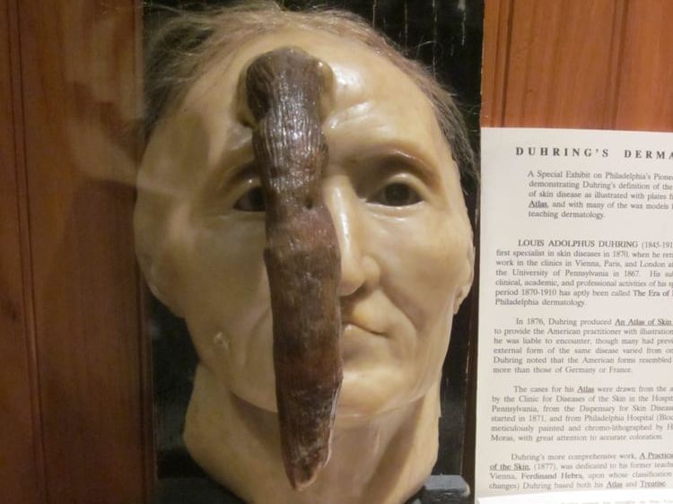
A chance encounter with what was to become his first well-known acquisition of a medical curiosity, Mutter started thinking on how to help those people that were known at that time as “monsters”, patients who the general public did not see, because they did not appear in public. The curiosity in question was a wax reproduction of the face of a French woman who had a “horn” arising from her forehead. This piece is on exhibit at the Mütter Museum.

Back in the United States in 1832, Thomas Dent Mutter changed his last name to give it a more “European” sound and added an “umlaut”, so now he was Thomas D. Mütter, MD. It may also be that he wanted to pay homage to his Scottish-German heritage, who knows? He opened his medical office in Philadelphia and although it took time, eventually he had a thriving practice. One of his specialties was the work on “deformities” so common at the time because of facial scars born out of the use of open fires in houses, and deformities caused by burns and loss of tissue due to chemicals used in local industry. Dr. Mütter is the pioneer of what we call today “Reconstructive Surgery”.

Wax curiosity, human horn
Wax figure, frontal horn

In 1835 he was asked to join the Medical Institute of Philadelphia as an assistant professor of Surgery. He was an instant success. Dr. Mütter was adored by his students because, he would question the students and guide them to discovery instead of just lecturing and leaving. In his Discourse eulogy of Dr. Mütter by Joseph Pancoast he writes:” The power of attracting students near him by his mingled gentleness, energy, and enthusiasm; of fixing their attention by the lucid and methodical arrangements of his Subject, by his clear demonstrations, and sprightly oral elucidations, came so readily to him, and was so early displayed) as to seem almost intuitive.” In 1841 Dr Mütter was appointed Professor of Surgery at the Jefferson Medical College in Philadelphia.

Dr. Mütter had always had poor health, even in childhood, and his dedication to his passion, long hours, took its toll on his body.

In 1956 he set sail for Europe and resigned his teaching duties. He was named Emeritus Professor of Surgery. Unfortunately, the trip did not help, and he returned to the US in early 1958. Fearful of another winter in cold Philadelphia, he moved to Charleston, SC, where he died on March 19, 1859.

Dr. Mütter’s story does not end here. He was an avid collector and throughout his short life he had pulled together an impressive collection of medical oddities, samples, and curiosities. Knowing that his life was at an end, he negotiated with the Philadelphia College of Physicians to have them host his collection in perpetuity as well as the creation of a trust fund that would ensure that the public and medical students would have access to this incredible collection. Through the years this collection has increased and is known today as the Mütter Museum of the Philadelphia College of Physicians. I strongly urge our readers to visit this incredible museum. For more information, click here.

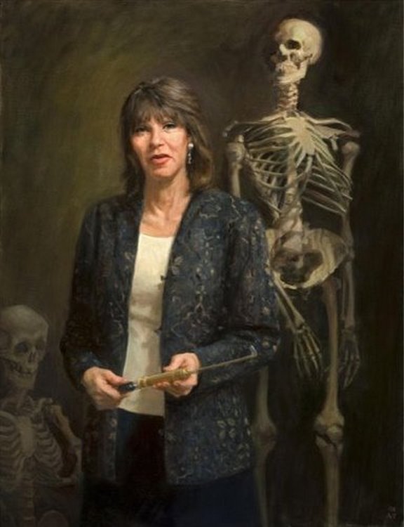
Personal notes: In the late 90’s, I attended a meeting of the American Association of Clinical Anatomists.  During the meeting I met Gretchen Worden, who at the time was the Curator of the Mütter museum. Gretchen was inspirational, fun, and a great conversationalist! I had the opportunity to visit Gretchen at the Mütter museum and had the luck to be treated to a “behind the scenes” tour. What an experience! I was saddened to hear that Gretchen Worden passed on August 2, 2004. Still, in my recent visit to the Mütter Museum, I was glad to see a new section at the museum that remembers Gretchen. Her biography can be read here.

I would like to thank Dr. Leslie Wolf for lending me the book by O’Keefe that lead to me writing this article. Dr. Miranda

Sources:

1. “Dr. Mütter’s Marvels: A True Tale of Intrigue and Innovation at the Dawn of Modern Medicine” O’Keefe, C. 2015 Penguin Random House, LLC
2. “A Discourse Commemorative of the Late Professor T.D. Mütter” Pancoast, J. 1859 J Wilson Publisher
3. “Thomas Dent Mütter: the humble narrative of a surgeon, teacher, and curious collector” Baker, J, et al. The American Surgeon, Atlanta 77:iss5 662-14 
4. “Thomas Dent Mutter, MD: early reparative surgeon” Harris, ES; Morgan, RF. Ann Plast Surg 1994 33(3):333-8 5.
5 "Things I Learned from Thomas Dent Mütter” O’Keefe C.
All images with permission from The Mutter Museum.


 IN MEMORIAM


Gretchen Worden (1947-2004)


Surface anatomy

Surface anatomy art by Danny Quirk - with permission

UPDATED: Surface anatomy is a subset of human anatomy that studies the relationship of external anatomical landmarks and deeply situated structures. These landmarks are created by cartilage, bones, tendons and muscles. In some cases they may be caused by a hypertrophic or diseased organ. Palpation is and art and my favorite book on the subject is the "Trail Guide to the Body" series by Andrew Biel

Surface anatomy is a core clinical skill for physicians, physical therapists and other health care professionals (HCP), as palpation in specific locations can lead to rapid diagnosis in certain cases.  It is also used in surgery to determine the appropriate place for incisions or insertion of a trocar in the case of minimally invasive surgery.

It is true that individual habitus and anatomical variations must make the HCP wary of potential mistakes, but in the majority of cases these anatomical landmarks and diagnoses are correct.

Many book chapters have been written on the topic, but unfortunately, the need to dedicate time to modern discoveries have reduced the time spent on this wonderful learning tool to the point that modern books of anatomy barely touch upon the subject. Many feel today that the need for this tool has been made irrelevant by the bed-side availability of ultrasound imaging.  I have to agree with Standring (2012) when she states “I am not convinced that surface anatomy is under growing threat from modern imaging technology…” There is and always will be the need for applied surface anatomy, although maybe not in the way and depth it was used in the past when imaging technology was scarce and expensive.

In order to make the importance of surface anatomy relevant in medical schools, some have added body painting as a tool in their anatomical curriculum with great results. Will it be used everywhere? I doubt it, but here again is a link between human anatomy and art, which our contributor Pascale Pollier presents through her art in "Artem Medicalis".

The artwork in this article is by Danny Quirk. Click on the image for a larger depiction. The video links are for the RMIT University Muscle Man and the Skeletal Man body paintings from the class by Dr. Claudia Diaz.

Personal note: One of the best book chapters on Surface Anatomy was written in "The Anatomical Basis of Clinical Practice" by Becker, Wilson and Gehweiler (1971). Unfortunately, although the content is very good, the images used, the tongue-in-cheek humor,  and writing style used by the authors, plus the times at which the book was published forced the publisher stop the sales of this book. It was banned from use in medical colleges through the country. Known to many as the "green book" because of its cover, it is today considered a collector's curiosity. In the "sources" section of this article you can find a links to a journal article on the subject as well as images of the book. Dr. Miranda.

Sources:
1. “Evidence-Based Surface Anatomy” Standrig, S J Clin Anat (2012) 25:813–815
2. “Giving Color to a New Curriculum: Body paint as a Tool in Medical Education” Den Akker, JW. Et al J Clin Anat (2002) 15:356–362
3. “Body-Painting: A Tool Which Can Be Used to Teach Surface Anatomy” Nanjundaiah, K. J Clin Diag Res (2012) October, Vol-6(8): 1405-1408 Copy of the article here
4. “RMIT students paint anatomical man into human textbook” RMIT Dr. Diaz, C. Copy of the article here
5. “Should We Use Body Painting to Teach Anatomy?” Gambino, M article at Smithsonian.com  
6. “The role of Fresh Tissue Dissection and Anatomic Body Painting in Anatomy Education” Bennet, C. PPT presentation on PDF here
7. " The anatomical Basis of Medical Practice" Becker, RF. Wilson, JW, Gehweiler, JA 1971, Baltimore, Williams & Wilkins
8. "The Pornographic Anatomy Book? The Curious Tale of The Anatomical Basis of Medical Practice" Halperin EC, Acad Med (2009) 84:2; 278-283 Article here
9. "The Objectification of Female Surface Anatomy" Ruiz, V. Internet article. Click here


Abdominal Regions

UPDATED: In surface anatomy the abdomen can be divided into nine regions by named lines (or planes): transpyloric, transtubercular, and midclavicular. These regions have specific visceral content.

• Hypochondriac regions (right and left): [Hypo]="below"; [chondr]="cartilage"; [iac]=”pertaining to”. In this context, the term means “below or deep to the cartilage (of the ribs)". The right hypochondriac region contains the liver, gallbladder, portal vein, and the right colic flexure. The left hypochondriac region contains the stomach, spleen, tail of the pancreas, and left colic flexure. For a detail on how the name of this region relates to a mental disorder, click here.

• Epigastric region: [Epi]="above"; [gastr]="stomach”; [ic]=”pertaining to". The term means “above the stomach”. This region contains mostly stomach and abdominal esophagus

• Lumbar regions (right and left): Right and left lumbar regions. The term [lumbar] refers to the area of the loins. The right lumbar region contains the ascending colon and part of the right kidney. The left lumbar region contains the descending colon and part of the left kidney

• Umbilical region: Centered around the umbilicus, this region contains mostly small intestine, abdominal aorta, and greater omentum


Abdominal regions - Modified from the original Davis, 1910 

• Inguinal regions (right and left): From the Latin [inguen]=”groin". Gaius Plinius Secundus aka “Pliny” (23-79 AD) first used this term naming after a plant (inguinalis) which he used to treat hernias of the groin. The right inguinal region contains the cecum and small intestine. The left inguinal region contains the sigmoid colon. In older days, as shown in the sketch, these regions were called the "iliac regions".

• Hypogastric region: [Hypo]="below"; [gastr]="stomach”; “ic”=”pertaining to. The term means “below the stomach”. The hypogastric region contains mostly small intestine and greater omentum. This region used to be called the "pubic region"

A clinical importance of these abdominal regions is that a ventral hernia is usually named by the anatomical region where it protrudes. Based on the image, you will see then why a hernia can be umbilical, inguinal, lumbar, epigastric, or hypogastric.

Sources:
1. "Clinical Anatomy" Brantigan, OC 1963 McGraw Hill
2. "Tratado de Anatomia Humana" Testut et Latarjet 8th Ed. 1931 Salvat Editores, Spain
3. Davis, Gwilym G. "Applied Anatomy: The Construction of the Human Body Considered in Relation to Its Functions, Diseases, and Injuries"; Philadelphia: J.B. Lippincott Co., 1910
Image modified from the original Davis, 1910

MTD Main Page Subscribe to MTD

Phrenoesophageal ligament

UPDATED: The [phrenoesophageal ligament] or phrenoesophageal membrane is part of a complex system that closes off the esophageal hiatus, one of the seven hiatuses in the respiratory diaphragm, preventing the herniation of abdominal structures into the thoracic mediastinum.

The connective tissue layer called the endoabdominopelvic fascia, which lines the inner aspect of the abdominopelvic cavity, is found as a "glue" between the respiratory diaphragm and the parietal peritoneum. At this point the endoabdominopelvic fascia is called the "infradiaphragmatic fascia".

When the infradiaphragmatic fasia gets to the edge of the esophageal hiatus, it splits into ascending and a descending components or limbs. These are the superior and inferior phrenoesophageal ligaments or phrenoesophageal membranes. These phrenoesophageal ligaments create a circular disc-like plug between the abdomen and the thorax. This "plug" is reinforced by a infradiaphragmatic fat pad found internal to the phrenoesophageal ligament.

The phrenoesophageal ligaments are reinforced externally. On their thoracic aspect by the endothoracic fascia, and on the abdominal side, by parietal peritoneum.

Esophageal hiatus
Images property of:CAA.Inc. Artist:Dr. E. Miranda
The ascending limb fuses superiorly with the esophageal fascia, which lines the external aspect of the longitudinal muscle of the esophagus, as the thoracic esophagus does not have a serosa layer. The descending limb fuses inferiorly with the esophageal fascial covering of the longitudinal ligament as it is covered by the peritoneum . Failure of the phrenoesophageal ligaments can predispose to esophageal hiatus hernia.
MTD Main Page Subscribe to MTD

2018 AACA Meeting

The following article is embedded from our Facebook page  https://www.facebook.com/CAAInc.

This year the 2018 meeting of the American Association of Clinical Anatomists is being held in Atlanta, GA., at the Grand Hyatt Buckhead Hotel and Conference Center. The program is full of interesting topics and is already a hit with all the attendees. Looking forward to the program.

2018AACAmeeting lg


MTD Main Page Subscribe to MTD