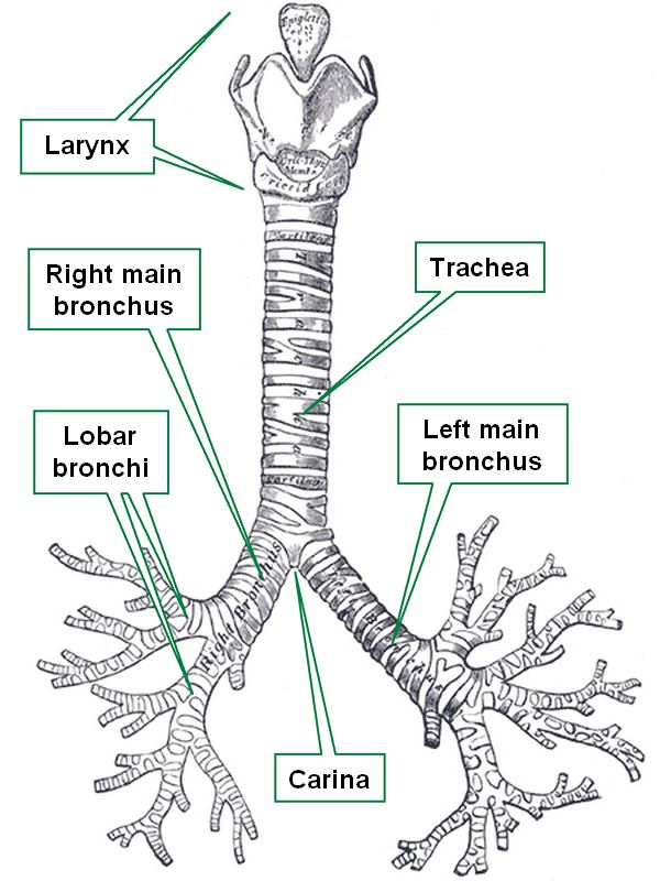
Tracheobronchial tree
The trachea, vernacularly known as the "windpipe", is in the adult a 4 inch (10.1 cm) membranous tube which contains incomplete, almost circular rings of cartilage. Some authors describe between fourteen to sixteen of these incomplete rings of cartilage in the trachea.
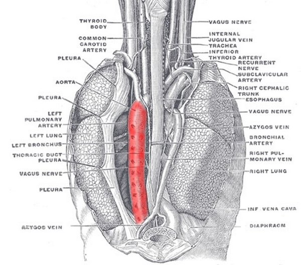
These semicircular rings are open-ended posteriorly, where the trachea only has a membrane, thus being flattened posteriorly. This posterior aspect of the trachea is in close anatomical relationship with the thoracic esophagus.
The trachea begins at the inferior end of the larynx, just about the level of the sixth cervical vertebra, and descends in the superior mediastinum to the level of the sternal angle (of Louis) close to the superior border of the fifth thoracic vertebra, where it divides into two main bronchi, each entering the ipsilateral lung at the level of the pulmonary hilum.
The word [trachea] arises from the Greek [trakhus] meaning "rough". This is because in the original description of this structure there was the belief that all arteries were full of air (Lat. air = ar/aria). The trachea, being rough was called the "rough artery" or "tracheartery", which was later amended, eliminating the "artery" part. In Latin the term used for the trachea was [arteria aspera], also meaning "rough artery".
The internal lower border of the bifurcation of the trachea into the left and right main bronchi is knows as the carina, where the carinal lymphatic nodes are found.
Sources:
1 "Tratado de Anatomia Humana" Testut et Latarjet 8 Ed. 1931 Salvat Editores, Spain
2. "Anatomy of the Human Body" Henry Gray 1918. Philadelphia: Lea & Febiger
Image modified by CAA, Inc. Original image courtesy of bartleby.com