Medical Terminology Daily (MTD) is a blog sponsored by Clinical Anatomy Associates, Inc. as a service to the medical community. We post anatomical, medical or surgical terms, their meaning and usage, as well as biographical notes on anatomists, surgeons, and researchers through the ages. Be warned that some of the images used depict human anatomical specimens.

You are welcome to submit questions and suggestions using our "Contact Us" form. The information on this blog follows the terms on our "Privacy and Security Statement" and cannot be construed as medical guidance or instructions for treatment.


We have 121 guests online


A Moment in History

Jean-Louis Petit

Jean Louis Petit
(1674 – 1750)

French surgeon and anatomist, Jean Louis Petit was born in Paris in on March 13, 1674.  His family rented an apartment at his house to Alexis Littre (1658 – 1726), a French anatomist. Petit became an apprentice of Littre at seven years of age, helping him in the dissections for his lectures and at an early age became the assistant in charge of the anatomic amphitheater.

Because of Petit’s dedication to anatomy and medicine, in 1690 at the age of sixteen, became a disciple of a famous Paris surgeon, Castel.

In 1692, Petit entered the French army and performed surgery in two military campaigns. By 1693 he started delivering lectures and was accepted as a great surgeon, being invited to the most difficult operations.  In 1700 he was appointed Chief Surgeon of the Military School in Paris and in the same year he received the degree of Master of Surgery from the Faculty of Paris.

In 1715 he was made a member of the Royal Academy of Sciences and an honorary member of the Royal Society of London. He was appointed by the King as the first Director General of the Royal Academy of Surgery when it was founded in 1731.

Petit’s written works are of historical importance.  “Traite des Maladies des Os” ( A Treatise on Bone Diseases);  “Traite des Maladies Chirurgicales et des Operation” (A Treatise on Surgical Diseases and their Operations” This last book was published posthumously in 1774. He also published a monograph on hemorrhage, another on lachrymal fistula, and others.

He was one of the first to perform choIecystotomy and mastoidotomy. His original tourniquet design for amputations saved many in the battlefield and the design of the same surgical instrument today has not changed much since its invention by him.

His name is remembered in the lumbar triangle, also called the "triangle of Petit", and the abdominal hernia that can ensue through that area of weakness, the lumbar hernia or "Petit's hernia".

Sources:
1. “Jean Louis Petit – A Sketch of his Life, Character, and Writings” Hayne, AP San Fran Western Lancet 1875 4: 446-454
2. “Oeuvres compl?tes de Jean-Louis Petit” 1837 Imprimerie de F. Chapoulaud
3. Extraits de l'eloge de Jean-Louis Petit Ius dans Ia seance publique de I' Academie royale de chirurgie du 26 mai 1750” Louis A. Chirurgie 2001: 126 : 475- 81


 "Clinical Anatomy Associates, Inc., and the contributors of "Medical Terminology Daily" wish to thank all individuals who donate their bodies and tissues for the advancement of education and research”.

Click here for more information


abebooks banner

bookplateink.com


Abraham Vater


This article is part of the series "A Moment in History" where we honor those who have contributed to the growth of medical knowledge in the areas of anatomy, medicine, surgery, and medical research.To search all the articles in this series, click here.

Abraham Vater (1684 - 1751) German anatomist and physician, Abraham Vater was the son of a distinguished physician and was born on the city of Wittenberg. He obtained a Doctorate in Philosophy (PhD) in 1706 and his medical degree in 1710 at the University of Leipzig. He became a professor “extraordinarius” of Anatomy and Botany in 1719, continuing his career in anatomy until he obtained the highest professorial degree at the University.

After the death of his father in 1733 Abraham Vater was appointed to the chair of Anatomy. In 1720 he published the discovery of a “biliary diverticulum” , the hepatopancreatic ampulla, known today as the “Ampulla of Vater” or duodenal papilla. His original article was entitled “Dissertatio anatomica qua novum bilis diverticulum circa orificium ductus choledochi”. He also described a large subcutaneous sensory nerve terminal known as the “Vater–Pacinian corpuscle”.

Known as an anatomist, Vater also wrote on surgery, gynecology, pharmacology, pathology, chemistry, and botany, making him a complete scientist. In a twist of fate, Vater died in 1751 after being afflicted with jaundice, probably a consequence of biliary blockage of his eponymic ampulla.

Abraham VaterOriginal image courtesy of www.nih.gov
Sources:
1. "Abraham Vater (1684-1751)" Brit Med J; 2,(4) (1951), 1214
2. "Abraham Vater of the Ampulla (Papilla) of Vater" Lerch, MM; Domschke, W. Gastroenterology (2000) 118: (2) 379
Back to MTD Main Page Subscribe to MTD

Epicardium

The word [epicardium] is composed by the prefix [epi-], meaning "outer" or "above"; the root term [-card-], meaning "heart"; and the suffix [-ium], meaning "layer" or "membrane". Thus, the word means "outer layer of the heart".

The epicardium is part of a larger structure called the pericardium, in fact, since the pericardium is in contact with a viscus (the heart), it can also be called "visceral pericardium". It must be noted that these two terms are synonyms: "epicardium" and "visceral pericardium".

The epicardium is a type of serosa composed of an outer layer formed by mesothelial cells and an inner layer formed by loose connective tissue containing varying amounts of fat. This inner layer known as the "subepicardial layer" (a misnomer) also contains elastic fibers.

HeartImage property of: CAA.Inc.Artist: Victoria G. Ratcliffe
The main arterial and venous components of the coronary circulation are found in the subepicardial layer. When performing a Coronary Artery Bypass Graft (CABG), a surgeon has to open the outer layer of the epicardium, and enter the subepicardial layer to perform the graft anastomosis

As a serosa, the epicardium is involved in the production and absorption of a clear fluid, the "pericardial fluid". This fluid acts as a lubricant for the pericardium, allowing for the effortless movement of the heart within the pericardial sac.

Back to MTD Main Page Subscribe to MTD

-iasis

The suffix [-iasis] has an original Greek root (such as in [iatros] meaning "healer" or "hospital"). It later became a Latin root meaning "condition, pathology, or disease".

This suffix can be found in many medical terms such as:

• Choledocolithiasis - a condition of stones in the bile duct.
• Elephantiasis - A condiction where a body part grows unusually large, usually lower extremities and/ or scrotum
• Phthiriasis- A condition or infestarion with public or crab lice
• Helminthiasis- Anbodily infestation with a type of parasitic worms called helminths (tapeworms)
• Cystolithiasis -  Bladder stones

Back to MTD Main Page Back to MTD Main Page

Pteryg / pter

Anterior view of the sphenoid bone. Public domain
Anterior view of the sphenoid bone


Both these root terms have their origin from the Greek [πτέρυγα] (ptéryga) and mean "wing".

In human anatomy the most common use of this root term is in the word [pterygoid]. Since the suffix [-oid] means "similar to", the word pterygoid means "similar to a wing", or "wing-like".

On the inferior aspect of the sphenoid bone (os sphenoidale) there are two very thin bat-wing-like bony appendages that are called the lateral and medial pterygoid plates. The medial pterygoid plate has a hook-like bony appendage called the hamulus (Latin: little hook).

Related to the pterygoid plates are the lateral and medial pterygoid muscles, both these muscles aid inthe process of mastication.

The root term [pter-] can be found in words such as [pterodactyl] meaning "winged finger", it refers to a phrehistoric winged animal; [pteranodon], and [pterosaurus].

Sources:
1 "Tratado de Anatomia Humana" Testut et Latarjet 8 Ed. 1931 Salvat Editores, Spain
2. "Anatomy of the Human Body" Henry Gray 1918. Philadelphia: Lea & Febiger
Image modified by CAA, Inc. Original image courtesy of bartleby.com

Note: Google Translate includes the symbol (?). Clicking on it will allow you to hear the pronunciation of the word.


Sinuatrial (SA) node

The sinuatrial node, is also known as the "sinoatrial node", "SA node" or by its eponym, the "node of Keith and Flack". It is the initial component of the conduction system of the heart.

It is a small nodule of cardiac muscle tissue, somewhat horseshoe-shaped that is found at the junction of the superior vena cava and the right atrium. Because of the inherent automaticity and rhythmical contractions of the SA node, it acts as the main pacemaker of the heart, being the base for normal heart beat, also known as "sinus rythm"

It receives blood supply from the SA node artery, usually the first or second branch that arises off the right coronary artery. The SA node artery is a long vessel that passes between the right atrium and the ascending aorta on its way to the SA node.

Conduction system of the heart

The SA node receives innervation from both sympathetic and parasympathetic nerves. The parasympathetic innervation is by way of the vagus nerve, the Xthcranial nerve. The sympathetic innervation is by way of the cardiac nerves, a plexus that has its origin in the ventral ramiof the first four thoracic spinal nerves (T1-T4).

The SA node was discovered in 1906 by Martin W.  Flack (1882-1931) and Arthur Keith (1866-1955). They named it originally the sinoauricular node.

Click on the image for a larger version. Image modified from the original: "3D Human Anatomy: Regional Edition DVD-ROM." Courtesy of Primal Pictures

Back to MTD Main Page Subscribe to MTD

Wilhelm His Jr.

This article is part of the series "A Moment in History" where we honor those who have contributed to the growth of medical knowledge in the areas of anatomy, medicine, surgery, and medical research.

Wilhelm His Jr.
Wilhelm His Jr.

Wilhelm His Jr. (1863-1934). Also known as Wilhelm His the Younger, was born in Switzerland in the city of Basel. His father was a well-known and famous anatomist by the same name who worked at the University of Basel. Wilhelm His Jr. studied in several universities, including Leipzig, Strasbourg, Bern, and Geneva. He received his medical degree from the University of Leipzig in 1889.

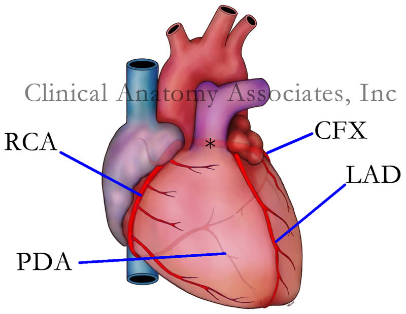
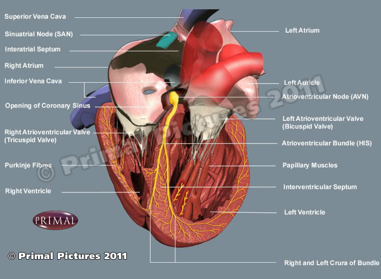
Although not an anatomist, his 1893 single and most brilliant contribution to the eventual understanding of the conduction system of the heart was the discovery and description of a muscular bundle that connected the atrial septum with the ventricular septum. Until that moment it was known that no muscles crossed from the atria to the ventricles, leading anatomists and physiologists to wonder how does the heart contract.  

In his article His states "after long search I have succeeded in finding a muscle bundle which unites the auricular and ventricular septal walls, and which, up to now, has escaped observation because of incomplete exposure, for it is visible in its entire extent only when the septa are cut exactly in their longitudinal direction".

What is interesting is that His not only found the bundle that today honors his name, but he included in that description the atrioventricular node, the actual structure that crosses the atrioventricular connective tissue barrier known as the "skeleton of the heart". The AV node was later to be clearly identified and researched by Sunao Tawara (1873 - 1952).

Dr. His continued his research on gout and joint diseases. He became a German citizen and joined the German army as a consulting physician. He retired in 1932 and died in 1934.

Although Wilhelm His Jr. did not discover or described the esophagogastric angle, in 1906 JD Cunningham started calling this angle the "Angle of His" in honor of Wilhelm His Jr. This eponym has stayed with us until today.

Sources:
1. "Wilhelm His Jr." JAMA. 1964;187(6):453-454
2. "His, Jr., W.: The Activity of the Embryonic Human Heart and Its Significance for the Understanding of the Heart Movement in the Adult" Arb Med Klin Leipzig, pp 14-49, 1893; Bast, TH,Gardner, WD trans: J Hist Med 4:289-318, 1949
3. "Wilhelm His, Jr. and the Bundle of His" Bast, TH, Gardner, WD J Hist Med All Sci; 1949; 4, (2) 170 -187
4. Firkin BG (1996) Dictionary of medical eponyms.London: Parthenon Publishing Group, 181
Original image
in the public domain, courtesy of the National Library of Medicine.