Medical Terminology Daily (MTD) is a blog sponsored by Clinical Anatomy Associates, Inc. as a service to the medical community. We post anatomical, medical or surgical terms, their meaning and usage, as well as biographical notes on anatomists, surgeons, and researchers through the ages. Be warned that some of the images used depict human anatomical specimens.

You are welcome to submit questions and suggestions using our "Contact Us" form. The information on this blog follows the terms on our "Privacy and Security Statement" and cannot be construed as medical guidance or instructions for treatment.


We have 103 guests online


A Moment in History

Jean-Louis Petit

Jean Louis Petit
(1674 – 1750)

French surgeon and anatomist, Jean Louis Petit was born in Paris in on March 13, 1674.  His family rented an apartment at his house to Alexis Littre (1658 – 1726), a French anatomist. Petit became an apprentice of Littre at seven years of age, helping him in the dissections for his lectures and at an early age became the assistant in charge of the anatomic amphitheater.

Because of Petit’s dedication to anatomy and medicine, in 1690 at the age of sixteen, became a disciple of a famous Paris surgeon, Castel.

In 1692, Petit entered the French army and performed surgery in two military campaigns. By 1693 he started delivering lectures and was accepted as a great surgeon, being invited to the most difficult operations.  In 1700 he was appointed Chief Surgeon of the Military School in Paris and in the same year he received the degree of Master of Surgery from the Faculty of Paris.

In 1715 he was made a member of the Royal Academy of Sciences and an honorary member of the Royal Society of London. He was appointed by the King as the first Director General of the Royal Academy of Surgery when it was founded in 1731.

Petit’s written works are of historical importance.  “Traite des Maladies des Os” ( A Treatise on Bone Diseases);  “Traite des Maladies Chirurgicales et des Operation” (A Treatise on Surgical Diseases and their Operations” This last book was published posthumously in 1774. He also published a monograph on hemorrhage, another on lachrymal fistula, and others.

He was one of the first to perform choIecystotomy and mastoidotomy. His original tourniquet design for amputations saved many in the battlefield and the design of the same surgical instrument today has not changed much since its invention by him.

His name is remembered in the lumbar triangle, also called the "triangle of Petit", and the abdominal hernia that can ensue through that area of weakness, the lumbar hernia or "Petit's hernia".

Sources:
1. “Jean Louis Petit – A Sketch of his Life, Character, and Writings” Hayne, AP San Fran Western Lancet 1875 4: 446-454
2. “Oeuvres compl?tes de Jean-Louis Petit” 1837 Imprimerie de F. Chapoulaud
3. Extraits de l'eloge de Jean-Louis Petit Ius dans Ia seance publique de I' Academie royale de chirurgie du 26 mai 1750” Louis A. Chirurgie 2001: 126 : 475- 81


 "Clinical Anatomy Associates, Inc., and the contributors of "Medical Terminology Daily" wish to thank all individuals who donate their bodies and tissues for the advancement of education and research”.

Click here for more information


abebooks banner

bookplateink.com


Respiratory Diaphragm

Respiratory diaphragm. Public domain
Respiratory diaphragm

The respiratory diaphragm is a musculotendinous dome-shaped shelf that divides the thoracic cavity from the abdominal cavity. It is one of the four named diaphragms in human anatomy. This is why it is proper to call it the "respiratory diaphragm" instead of just "diaphragm".

The name comes from the Greek, where the prefix [dia-] means "complete" or "through" and [phragm] means "a partition". In Latin, Celsus called it the "septum transversum", meaning "the transverse partition". The root term for [diaphragm] is [-phren-], as in phrenic nerves and cardiacophrenic vessels.

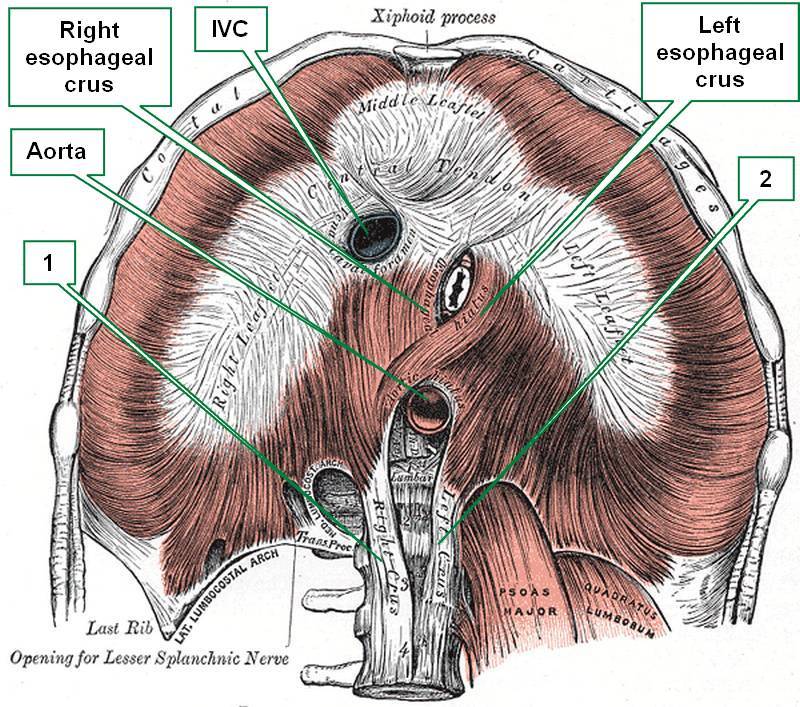
The respiratory diaphragm has a club-shaped central tendon (as in a card suit) which has a large hiatus for the inferior vena cava. The muscular portion is formed by skeletal (voluntary) muscle and descends skirt-like to attach to the internal aspect of the sternum and ribs anterolaterally, and a complex system of lumbocostal tendinous arches posteriorly.

It receives its blood supply through branches of the intercostal arteries, the musculophrenic artery, and the pericardiacophrenic arteries. It is innervated by the phrenic nerves, which descend through the mediastinum in relation to the pericardium.

The respiratory diaphragm has not one, but seven openings (hiatuses) to allow for passageway of many structures:

• Esophageal hiatus
• Aortic hiatus
• Inferior vena cava hiatus
• Hiatuses (2) for the superior epigastric vessels, which are the inferior continuation of the internal thoracic (mammary) vessels. Also known as the hiatuses of Morgagni.
• Hiatuses (2) for the splanchnic nerves

The most common site for an internal abdominothoracic herniation is the esophageal hiatus, but there are other respiratory diaphragm hernias, including the retrosternal hernia of Morgagni (through the superior epigastric hiatus), and the hernia of Bochdalek (a congenital hernia through an incomplete central tendon of the respiratory diaphragm).

Sources:
1 "Tratado de Anatomia Humana" Testut et Latarjet 8 Ed. 1931 Salvat Editores, Spain
2. "Anatomy of the Human Body" Henry Gray 1918. Philadelphia: Lea & Febiger Image modified by CAA, Inc. Original image by Henry Vandyke Carter, MD., courtesy of bartleby.com


Jejunum

<span>Jejunoileal vascular supply</span><span></span>. Public domain
Jejunoileal vascular supply

The jejunum is an intraperitoneal organ, it is the second portion of the small intestine and part of the digestive tract. It begins at the duodenojejunal junction  where it is related to the ligament of Treitz, and extends 8 to 9 feet, continuing distally with the ileum.

Being intraperitoneal, it is anchored to the posterior abdominal wall by the double-layered mesentery through which the jejunum receives its blood and nerve supply. At the root (base) of the mesentery are the superior mesenteric vessels.

The Latin word [jejunis] means "empty" or "fasting". The Latin term [jejunum] was used by the Romans to denote the first meal of the day, breakfast, when you have an "empty" stomach. The term was associated with this segment of the small intestine, as it is most of the time found empty in cadavers being dissected.

There is no clear anatomical boundary between the jejunum and ileum, as they blend smoothly one into the other. There are several gross changes from jejunum to ileum, one of them being that the complexity of the mesenteric arterial arches increases from proximal to distal. See the accompanying image. Click on it for a larger depiction.

Two interesting side notes: In English, the term for the first meal of the day is self-explanatory: [break - fast], adding to the Roman concept of "fasting" or "jejunum". In Spanish, the term for breakfast is [desayuno], where the word [ayuno] means "fasting", therefore the word [des-ayuno] also means "the end of fasting". Look at the evolution (in Spanish) from [jejunum] to [yeyuno] (the Spanish term for the organ) to [ayuno], meaning "fasting" or "empty".

Sources:
1. "The Origin of Medical Terms" Skinner, HA 1970 Hafner Publishing Co.
2. "Clinically Oriented Anatomy" Moore, KL. 3r Ed. Williams & Wilkins 1992
3 "Tratado de Anatomia Humana" Testut et Latarjet 8 Ed. 1931 Salvat Editores, Spain
4. "Anatomy of the Human Body" Henry Gray 1918. Philadelphia: Lea & Febiger Image modified by CAA, Inc. Original image by Henry Vandyke Carter, MD., courtesy of bartleby.com


Crus / crura

Respiratory diaphragm - anteroinferior view (modified from bartleby.com) 1. Right tendinous aortic crus 2. Left tendinous aortic crus IVC=Inferior Vena Cava
1. Right tendinous aortic crus 2. Left tendinous aortic crus
IVC= Inferior vena cava

The word [crus] is Latin (cruris) and refers to the leg, or region of the shin. It is commonly used to mean "leg" or "pillar". The plural form is [crura].

Several authors suggest a relation of [crus] with another Latin term [crux] meaning "cross" as if a cross is formed by two [crura] (legs).

The term crus is widely used in human anatomy:

- crus cerebri: there are two crus cerebri in the anterior aspect of the mesencephalon
- crura of the penis: the posterior aspect of the corpora cavernosa firmly attached to the ischiopubic rami
- crura of the clitoris:  the posterior aspect of the corpora cavernosa firmly attached to the ischiopubic rami
- crura fornix cerebrii: the posterior converging bands that form the fornix of the cerebrum

Special mention is deserved by the crura of the diaphragm. There are two pairs of diaphragmatic crura. The esophageal crura (right and left) which bound the passageway of the esophagus from the thorax into the abdomen, the esophageal hiatus. The esophageal crura have a muscular structure. The aortic crura (righ and left) allow for passage of the aorta into the abdomen, and although muscular superiorly, they are mostly tendinous. The accompanying image shows an anteroinferior view of the respiratory diaphragm. Click on the image for a larger picture.

Sources:
1. "The Origin of Medical Terms" Skinner, HA 1970 Hafner Publishing Co.
2. "Medical Meanings - A Glossary of Word Origins" Haubrich, WD. ACP Philadelphia
3 "Tratado de Anatomia Humana" Testut et Latarjet 8 Ed. 1931 Salvat Editores, Spain
4. "Anatomy of the Human Body" Henry Gray 1918. Philadelphia: Lea & Febiger
Image modified by CAA, Inc. Original image by Henry Vandyke Carter, MD., courtesy of bartleby.com


Luigi A. Galvani


This article is part of the series "A Moment in History" where we honor those who have contributed to the growth of medical knowledge in the areas of anatomy, medicine, surgery, and medical research.To search all the articles in this series, click here.

Luigi A. Galvani (1737-1798). Italian anatomist, surgeon, and physiologist, Luigi Aloisio Galvani was born in Bologna in 1737. Although he started his studies to join the church, Galvani followed with medical studies at the University of Bologna, where he became a skilled anatomist and surgeon. On July 15, 1759 Galvani obtained his degree in medicine and philosophy.

He was interested in the effects of electricity on tissues and through observation and experimentation he postulated the existence of "animal electricity", that is, electricity generated within the tissues. He postulated the possibility that nerves carried electricity. His theories led to a passionate controversy with Volta, who denied Galvani's postulates. Galvani's theories would only be confirmed after his death. 

Galvani was deeply religious, and when forced by government officials to take an oath of atheism, he refused. He was stripped of his position and was lead to poverty. His position was restored close to his death. In his honor, Andre Ampere (1775-1836) named one of his inventions that measures electricity,  the "galvanometer". His name is also present in vernacular English, when we say that a rock star or a movie "galvanizes" an audience, meaning it was "electrifying"!

Sources:
1. "Luigi Galvani" Haas LF J Neurol Neurosurg Psychiatry v.56(10); Oct 1993
2. "Luigi Galvani and the foundations of electrophysiology" Cajavilca C, Varonb,J,Sternbachc GL; Resuscitation 80 (2009) 159–162

Luigi Galvani

Original imagecourtesy of National Institutes of Health.

Back to MTD Main Page Subscribe to MTD

Capitate

The capitate bone is one of the four bones that comprise the distal row of the carpus or carpal bones that form the wrist. It is the largest of the carpal bones and is placed in the center of the wrist (see image).

Its name originates from the Latin [caput], meaning "head". The capitate bone presents a large, rounded area, called the "head". To complete the homology, the capitate bone also has a narrow segment called the "neck", the rest of the bone called the "body". It is also known as "os capitatum" or "os magnum"

The capitate bone articulates with seven bones, including the scaphoid, lunate, trapezoid, hamate, and the three central metacarpals (2nd, 3rd, and 4th).

The accompanying image shows the anterior (volar) surface of the wrist. Click on the image for a larger picture.

Scaphoid bone - anterior (volar) view of the wrist

Image modified from the original: "3D Human Anatomy: Regional Edition DVD-ROM." Courtesy of Primal Pictures

Back to MTD Main Page Subscribe to MTD

Duodenum

The duodenum is a mostly retroperitoneal organ, part of the digestive tract, and the most proximal portion of the small intestine. This organ is approximately 10 inches in length (24.5 cm). It starts at the pylorus of the stomach, has a "C" shape, curving around the head and the neck of the pancreas, to end at the duodenojejunal junction.

The duodenum is described as having four segments of differing length, usually named numerically:

- First segment: about two inches in length, it is dilated and called the "duodenal ampulla", or "superior duodenum"
- Second segment: about three inches in length, it receives bile and pancreatic juice through the hepatopancreatic ducts and ampullae. It is also called the "descending duodenum"
- Third segment: about four inches in length, it crosses the midline, and is also known as the "horizontal" or "transverse duodenum"
- Fourth segment: one inch in length, this is the shortest segment, it ascends towards the duodenojejunal junction, which is tethered to the diaphragm by a fold of peritoneum around a fibromuscular band called the "ligament of Treitz". At this point the retroperitoneal duodenum becomes the intraperitoneal jejunum. This fourth segment is also called the "ascending duodenum"

Retroperitoneal organsImage property of:CAA.Inc.Artist:Dr. E. Miranda
The name of the organ is interesting. Most textbooks claim that is originates from the Latin [duodeni], meaning "twelve". The fact is that the duodenum was originally named in Greek [δώδεκα δάχτυλαν] meaning "twelve fingers". If you place both your hands together and add 1/4 of an inch to each side (as if you had an extra finger on each hand) that measures approximately 10 inches. The term was shortened by an incorrect translation to "twelve" by Gerard of Cremona (1114 - 1187) who called it "duodenum", a bad translation, as twelve fingers in Latin is [duodecim digitorum].

Sources:
1.
"Clinically Oriented Anatomy" Moore, KL. 3r Ed. Williams & Wilkins 1992
2. "The origin of Medical Terms" Skinner, AH, 1970 

Back to MTD Main Page Subscribe to MTD