Medical Terminology Daily (MTD) is a blog sponsored by Clinical Anatomy Associates, Inc. as a service to the medical community. We post anatomical, medical or surgical terms, their meaning and usage, as well as biographical notes on anatomists, surgeons, and researchers through the ages. Be warned that some of the images used depict human anatomical specimens.

You are welcome to submit questions and suggestions using our "Contact Us" form. The information on this blog follows the terms on our "Privacy and Security Statement" and cannot be construed as medical guidance or instructions for treatment.


We have 160 guests online


A Moment in History

Jean-Louis Petit

Jean Louis Petit
(1674 – 1750)

French surgeon and anatomist, Jean Louis Petit was born in Paris in on March 13, 1674.  His family rented an apartment at his house to Alexis Littre (1658 – 1726), a French anatomist. Petit became an apprentice of Littre at seven years of age, helping him in the dissections for his lectures and at an early age became the assistant in charge of the anatomic amphitheater.

Because of Petit’s dedication to anatomy and medicine, in 1690 at the age of sixteen, became a disciple of a famous Paris surgeon, Castel.

In 1692, Petit entered the French army and performed surgery in two military campaigns. By 1693 he started delivering lectures and was accepted as a great surgeon, being invited to the most difficult operations.  In 1700 he was appointed Chief Surgeon of the Military School in Paris and in the same year he received the degree of Master of Surgery from the Faculty of Paris.

In 1715 he was made a member of the Royal Academy of Sciences and an honorary member of the Royal Society of London. He was appointed by the King as the first Director General of the Royal Academy of Surgery when it was founded in 1731.

Petit’s written works are of historical importance.  “Traite des Maladies des Os” ( A Treatise on Bone Diseases);  “Traite des Maladies Chirurgicales et des Operation” (A Treatise on Surgical Diseases and their Operations” This last book was published posthumously in 1774. He also published a monograph on hemorrhage, another on lachrymal fistula, and others.

He was one of the first to perform choIecystotomy and mastoidotomy. His original tourniquet design for amputations saved many in the battlefield and the design of the same surgical instrument today has not changed much since its invention by him.

His name is remembered in the lumbar triangle, also called the "triangle of Petit", and the abdominal hernia that can ensue through that area of weakness, the lumbar hernia or "Petit's hernia".

Sources:
1. “Jean Louis Petit – A Sketch of his Life, Character, and Writings” Hayne, AP San Fran Western Lancet 1875 4: 446-454
2. “Oeuvres compl?tes de Jean-Louis Petit” 1837 Imprimerie de F. Chapoulaud
3. Extraits de l'eloge de Jean-Louis Petit Ius dans Ia seance publique de I' Academie royale de chirurgie du 26 mai 1750” Louis A. Chirurgie 2001: 126 : 475- 81


 "Clinical Anatomy Associates, Inc., and the contributors of "Medical Terminology Daily" wish to thank all individuals who donate their bodies and tissues for the advancement of education and research”.

Click here for more information


abebooks banner

bookplateink.com


Lumbar vertebra

The lumbar region of the spine is composed by five lumbar vertebrae. Although sharing characteristics common to all vertebrae, a lumbar vertebra can be differentiated by the following:

• The vertebral body is large, tall, and when viewed from superior, the vertebral body has a longer transverse diameter and a shorter anteroposterior diameter. Most anatomists describe the vertebral body as being “kidney-shaped”. The massiveness of the vertebral body is due to the larger weight that each consecutive vertebra has to bear in the lumbar region. Because of this, lumbar vertebrae have larger anular epiphyses, and the vertebral body is hourglass-shaped with a “waist”.  As with other vertebrae, the body of a lumbar vertebra has vertebral endplates, nutritional foramina, and in its posterior aspect they present with basivertebral foramina.

• The pedicles in the lumbar vertebrae are larger, course in a mostly anteroposterior direction, and have a oval cross-section where the superoinferior diameter is longer and the transverse diameter is shorter. This is important for transpedicular procedures for vertebroplasty or kyphoplasty.

• The transverse processes are also larger than other vertebrae. Of interest are the facts that the transverse process of the third lumbar vertebra is the longest of all lumbar vertebrae and that the transverse process of the fifth lumbar vertebra courses superolaterally at an angle of  almost 45 degrees because of the presence of the strong iliolumbar ligament. The lumbar transverse processes have a small tubercle called the mammillary process.

• The lumbar spinous processes are large and have a square shape.

Again, because of the larger weight bearing capacity of the lumbar vertebrae, they have strong ligaments and their zygapophyseal joints and articular facets tend to be oriented with their surfaces in a transverse plane. This has two consequences: The rotational mobility of the whole lumbovertebral region is limited and they tend not to have anteroposterior displacement.

Lumbar vertebra - lateral view
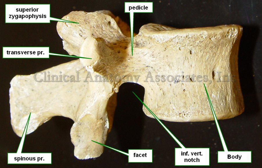
Lumbar vertebra - Lateral view.

Lumbar vertebra - inferior view
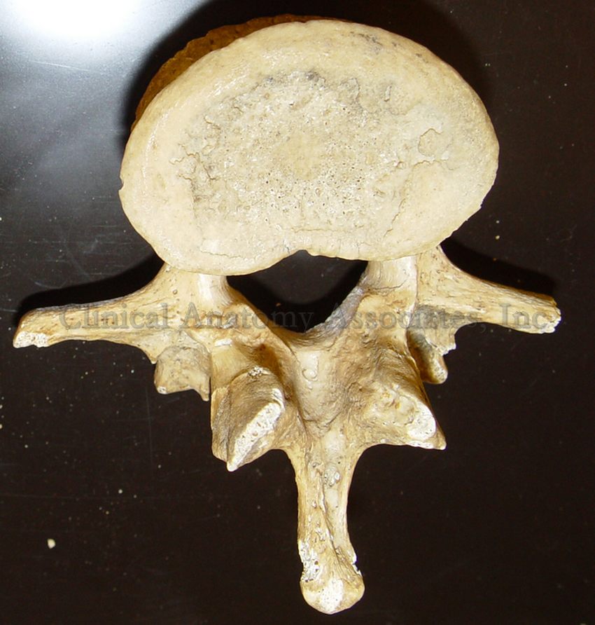
Lumbar vertebra - Inferior view.

Image property of CAA.Inc. Photographer:D.M. Klein.

Back to MTD Main Page Back to MTD Main Page