Medical Terminology Daily (MTD) is a blog sponsored by Clinical Anatomy Associates, Inc. as a service to the medical community. We post anatomical, medical or surgical terms, their meaning and usage, as well as biographical notes on anatomists, surgeons, and researchers through the ages. Be warned that some of the images used depict human anatomical specimens.

You are welcome to submit questions and suggestions using our "Contact Us" form. The information on this blog follows the terms on our "Privacy and Security Statement" and cannot be construed as medical guidance or instructions for treatment.


We have 96 guests online


A Moment in History

Jean-Louis Petit

Jean Louis Petit
(1674 – 1750)

French surgeon and anatomist, Jean Louis Petit was born in Paris in on March 13, 1674.  His family rented an apartment at his house to Alexis Littre (1658 – 1726), a French anatomist. Petit became an apprentice of Littre at seven years of age, helping him in the dissections for his lectures and at an early age became the assistant in charge of the anatomic amphitheater.

Because of Petit’s dedication to anatomy and medicine, in 1690 at the age of sixteen, became a disciple of a famous Paris surgeon, Castel.

In 1692, Petit entered the French army and performed surgery in two military campaigns. By 1693 he started delivering lectures and was accepted as a great surgeon, being invited to the most difficult operations.  In 1700 he was appointed Chief Surgeon of the Military School in Paris and in the same year he received the degree of Master of Surgery from the Faculty of Paris.

In 1715 he was made a member of the Royal Academy of Sciences and an honorary member of the Royal Society of London. He was appointed by the King as the first Director General of the Royal Academy of Surgery when it was founded in 1731.

Petit’s written works are of historical importance.  “Traite des Maladies des Os” ( A Treatise on Bone Diseases);  “Traite des Maladies Chirurgicales et des Operation” (A Treatise on Surgical Diseases and their Operations” This last book was published posthumously in 1774. He also published a monograph on hemorrhage, another on lachrymal fistula, and others.

He was one of the first to perform choIecystotomy and mastoidotomy. His original tourniquet design for amputations saved many in the battlefield and the design of the same surgical instrument today has not changed much since its invention by him.

His name is remembered in the lumbar triangle, also called the "triangle of Petit", and the abdominal hernia that can ensue through that area of weakness, the lumbar hernia or "Petit's hernia".

Sources:
1. “Jean Louis Petit – A Sketch of his Life, Character, and Writings” Hayne, AP San Fran Western Lancet 1875 4: 446-454
2. “Oeuvres compl?tes de Jean-Louis Petit” 1837 Imprimerie de F. Chapoulaud
3. Extraits de l'eloge de Jean-Louis Petit Ius dans Ia seance publique de I' Academie royale de chirurgie du 26 mai 1750” Louis A. Chirurgie 2001: 126 : 475- 81


 "Clinical Anatomy Associates, Inc., and the contributors of "Medical Terminology Daily" wish to thank all individuals who donate their bodies and tissues for the advancement of education and research”.

Click here for more information


abebooks banner

bookplateink.com


Right coronary artery

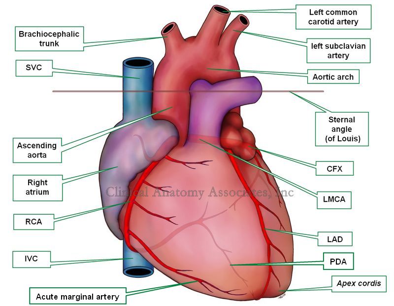
The right coronary artery (RCA) is one of the two branches that arises from the ascending aorta and provide blood supply to the heart. The RCA begins at the coronary ostium, situated usually within the right sinus of Valsalva found in the aortic valve, one of the semilunar valves of the heart.

The RCA gives off in its initial course two arteries: the conus artery, which gives blood to the conus arteriosus, the outflow tract of the right ventricle, and the artery to the sinuatrial (SA) node, a component of the conduction system of the heart.

The RCA descends in the atrioventricular sulcus, giving off a series of small right ventricular branches and a couple of small right atrial branches, it then bends around the acute margin (margo acutus) and passes to the posterior surface of the heart. Just before the RCA bends posteriorly, it will give off the acute marginal artery, usually a thin, longer branch that extends towards the cardiac apex.

Heart, anterior view. SVC: superior vena cava. RCA: right coronary artery; IVC: inferior vena cava; CFX: circumflex artery; LMCA: left main coronary artery; LAD: left anterior descending artery

Heart - Anterior view. Click on the image for a larger version.

In its posterior trajectory the RCA gives off a couple of small posterior right ventricular arteries and then ends at the crux cordis, where the RCA gives off the posterior interventricular artery, commonly known as the posterior descending artery (PDA). The RCA will also give off the posterolateral artery, which, situated in the atrioventricular sulcus, extends the vascular territory of the RCA into the region of the left ventricle. This origin of the PDA from the RCA is subject to anatomical variation, which gives origin to the concept of coronary dominance.

Arising from the terminal portion of the RCA (sometimes from the posterolateral artery) is the artery to the atrioventricular (AV) node, another component of the conduction system of the heart. It is easily understood that stricture or stenosis of the RCA (depending on location) can then lead to damage of the conduction system of the heart.

Human heart and coronary artery anatomy and pathology are some of the many lecture topics developed and presented by Clinical Anatomy Associates, Inc.

Image property of:CAA.Inc.Artist:Victoria G. Ratcliffe

Back to MTD Main Page Subscribe to MTD