Medical Terminology Daily (MTD) is a blog sponsored by Clinical Anatomy Associates, Inc. as a service to the medical community. We post anatomical, medical or surgical terms, their meaning and usage, as well as biographical notes on anatomists, surgeons, and researchers through the ages. Be warned that some of the images used depict human anatomical specimens.

You are welcome to submit questions and suggestions using our "Contact Us" form. The information on this blog follows the terms on our "Privacy and Security Statement" and cannot be construed as medical guidance or instructions for treatment.


We have 117 guests online


A Moment in History

Jean-Louis Petit

Jean Louis Petit
(1674 – 1750)

French surgeon and anatomist, Jean Louis Petit was born in Paris in on March 13, 1674.  His family rented an apartment at his house to Alexis Littre (1658 – 1726), a French anatomist. Petit became an apprentice of Littre at seven years of age, helping him in the dissections for his lectures and at an early age became the assistant in charge of the anatomic amphitheater.

Because of Petit’s dedication to anatomy and medicine, in 1690 at the age of sixteen, became a disciple of a famous Paris surgeon, Castel.

In 1692, Petit entered the French army and performed surgery in two military campaigns. By 1693 he started delivering lectures and was accepted as a great surgeon, being invited to the most difficult operations.  In 1700 he was appointed Chief Surgeon of the Military School in Paris and in the same year he received the degree of Master of Surgery from the Faculty of Paris.

In 1715 he was made a member of the Royal Academy of Sciences and an honorary member of the Royal Society of London. He was appointed by the King as the first Director General of the Royal Academy of Surgery when it was founded in 1731.

Petit’s written works are of historical importance.  “Traite des Maladies des Os” ( A Treatise on Bone Diseases);  “Traite des Maladies Chirurgicales et des Operation” (A Treatise on Surgical Diseases and their Operations” This last book was published posthumously in 1774. He also published a monograph on hemorrhage, another on lachrymal fistula, and others.

He was one of the first to perform choIecystotomy and mastoidotomy. His original tourniquet design for amputations saved many in the battlefield and the design of the same surgical instrument today has not changed much since its invention by him.

His name is remembered in the lumbar triangle, also called the "triangle of Petit", and the abdominal hernia that can ensue through that area of weakness, the lumbar hernia or "Petit's hernia".

Sources:
1. “Jean Louis Petit – A Sketch of his Life, Character, and Writings” Hayne, AP San Fran Western Lancet 1875 4: 446-454
2. “Oeuvres compl?tes de Jean-Louis Petit” 1837 Imprimerie de F. Chapoulaud
3. Extraits de l'eloge de Jean-Louis Petit Ius dans Ia seance publique de I' Academie royale de chirurgie du 26 mai 1750” Louis A. Chirurgie 2001: 126 : 475- 81


 "Clinical Anatomy Associates, Inc., and the contributors of "Medical Terminology Daily" wish to thank all individuals who donate their bodies and tissues for the advancement of education and research”.

Click here for more information


abebooks banner

bookplateink.com


Atrial fibrillation

EKG - Atrial Fibrillation, courtesy Dr. Randall K Wolf
Atrial fibrillation EKG

[UPDATED] What is atrial fibrillation?

Atrial fibrillation (AFib) is one of the most common heart conditions, affecting 4% of the adult population. Characterized by a rapid, irregular heartbeat, AFib is largely due to abnormal electrical impulses that cause the atria of the heart to quiver when it should be beating steadily.The atria are the two upper chambers of the heart.

Because of this quivering action, blood flow is reduced and is not completely pumped out of the atria.  This negatively impacts cardiac performance and also allows the blood to pool and potentially clot. These clots, if freed, can enter the systemic circulation and cause a stroke.

At rest, a normal heart rate is approximately 60 – 100 beats per minute.  In a person with AFib, that heart rate can increase to 180 bpm or even higher.  Thorough testing by your health care provider can spot abnormalities in the heart's rhythm before any obvious symptoms are noticed.

What are the symptoms?

Whether it is caused by stress, exercise, or too much caffeine, most people experience rapid heart from time to time.  Most cases are harmless, but AFib is a serious medical condition that may often be long lasting.  Some people with AFib experience no symptoms at all.  But for others,  AFib may cause:

    Exercise intolerance
    Fatigue
    Severe shortness of breath
    Chest pain
    Palpitations
    Light-headiness

What causes atrial fibrillation?

Your heart is divided into four chambers: the two upper chambers called atria, and two lower chambers called ventricles. In order for blood to be pumped through your body, a group of specialized cardiac cells, the conduction system of the heart,  sends electrical impulses to the atria that tells your heart to contract. Contractions of the heart send approximately five quarts of blood through your body every minute. In people with AFib, however, the impulses are sent chaotically. The atria quiver instead of beat; the blood isn't completely pumped out and may pool and potentially clot. AFib is a leading cause of stroke because of the anatomy of the left ventricle. For more information, read this article.

Are you at risk?

Your chances of developing AFib increase with age.  AFib occurs more commonly in women than in men.  According to the Framingham Heart Study, AFib is associated with a higher risk of death for women than for men. You are also at greater risk of developing AFib if you suffer from an overactive thyroid, high blood pressure, a prior heart attack, congestive heart failure, valve disease, or congenital disorders.

Diagnosis

AFib can sometimes be diagnosed with a stethoscope during an exam by a doctor or other health care provider and is confirmed or diagnosed with an electrocardiogram (EKG). There are several types of EKG’s. They are:

Resting EKG – Electrical activity in the heart is monitored when a person is at rest.
Exercise EKG – Activity is monitored when a person jogs on a treadmill or exercises on a stationary bike.
24-hour EKG (Holter Monitor) – A person wears a small, portable monitor that detects activity over the course of a day.
Transtelephonic event monitoring – A person wears a monitor for a period of a few days to several weeks. When AF is felt, the person telephones a monitoring station or activates the monitor's memory function. This type of EKG is particularly useful in detecting AF that occurs only once every few days or weeks. Unfortunately this type of monitor does not record heart events while you are sleeping.

The image on this article is a typical EKG AFib recording showing the flutter of the atria followed by the ventricular contraction. In the larger image (click on the image of the article) you can see how this fluttering of the atria causes an abnormal spacing of the ventricular contractions which some patients feel in their chest.

PERSONAL NOTE: For more information on AFib and its surgical treatment, click here.

Thanks toDr. Randall Wolf for the image andlinks


2023 Vesalius Triennial AEIMS, Antwerp – Belgium

Dr. Miranda speaking at the 2023 Vesalius Triennial
Dr. Miranda speaking at the
2023 Vesalius Triennial

I had the honor of being invited by the University of Antwerp in Belgium to speak at the 2023 Vesalius Triennial Meeting in the city of Antwerp. This scientific meeting was presented in conjunction with the 29th Congress of the Association Européenne des Illustrateurs Médicaux et Scientifiques - AEIMS (European Association of Medical and Scientific Illustrators). A three-day program that, alongside the scientific program, included poetry, art, music, sculpture, and painting. All of this celebrating the life and inspiration brought to arts and medical science by Andreas Vesalius Bruxellensis (1514-1564).

The scientific program included lectures by well-known Vesalius scholars, including Vivian Nutton, Robrecht Van Hee, Francis Van Glabbeek, Philip Van Kerrebroeck, Omer Steeno, Maurits Beisbrouck, Theodor Godeeris, Peter Bols, and many others. Personally, it was incredible to be invited to this event and share with these individuals.

One of the events of this meeting was an afternoon concert entitled “Vesalian landscapes in music, poetry, and photographs” by pianist Elke Robersscheuten, and my friend Theo Dirix, who read the poetry. This was accompanied by slides of Vesalian works, and images of the city of Brussels and the island of Zakynthos, Greece. One of the pieces performed by Elke Robersscheuten was “André Vésale”. Ths rare piece of piano sheet music is the topic for a separate article in this blog: An anatomical surprise from a French composer.

My presentation was entitled “Vesalius and Anatomical Megadrawings – A Personal Journey”. This is a topic that touched on my experience with larger (and very small) books and the sentence written by Andreas Vesalius in the two-page letter to Johannes Oporinus printed in the first part of Vesalius’ opus magnum “De Humani Corporis Fabrica, Libri Septem”. Referring to anatomical images, Vesalius states “quod tabulas quæe nunquam satis magna studiosis proponi poterunt”. Daniel H. Garrison in the latest publication of the Fabrica translates this as “illustrations which could never be large enough for students”.

The need for better resolution and the limitation of the printing technology (hand-carved woodblocks) at the time as well as the quality of the paper available forced the need for larger images. The Fabrica is a folio-size book and the images for the first time are labeled with letters, symbols, and characters with a detailed key as to their meaning.

The research for this presentation took me to the largest anatomy book ever printed, the “Anatomiæ Universæ Icones” by Giovanni Paolo Mascagni (1755 – 1815), a double elephant folio size book measuring 40 by 30 inches with two sets of 44 plates. This book was printed in black and white and hand-colored by Antonio Serantoni (1780 – 1837), an Italian engraver and painter. The printing and coloring of this book took ten years between 1823 and 1832. An incredible book of which there are 16 known copies in the world, one of them at the University of Cincinnati.

Anatomiæ Universæ Icones by Paolo Mascagni at the University of Cincinnati

One of the most interesting aspects of this book, besides the large size of each image, is the fact that a 5.9 feet tall human can be constructed if three pages are cut and placed together. Of course, this cannot be done with these incredibly rare and expensive books; but digital technology allows us to scan and lightly correct the background to eliminate imperfections and damage caused by 200 years of use.

With the help of the University of Cincinnati authorities, Gino Pasi (archivist and curator of the Henry R. Winkler Center for the History of the Health Professions at UC), and Samantha Scheck (graphic designer) we were able to access the Mascagni book, measure the images, scan them, and them digitally process them. The result were two large images that I took to Antwerp, receiving incredible feedback from the attendees.

Working with the Mascagni book

The life-size anatomy imagined by Mascagni

There is so much more to the life of Paolo Mascagni, before and after his death that include prison, family problems, greed, plagiarism, and a separate individual that is now known for his “dubious character”. I will write separate articles on these topics.

My presentation also touched on the large poster-like drawings (not books) that were used for anatomical teaching before the advent of the 35 mm slide projector and later PowerPoint with halogen light bulb projectors and today large LED monitors.

The anatomy amphitheater at the University of Chile Medical School

3 by 4 feet scroll painting by Juan Frutos M.

My alma mater, the University of Chile Medical School,has a museum and an old wooden amphitheater where I studied anatomy many years ago. As seen in the accompanying image, this auditorium has two incredibly large hand-drawn images that measure 13 feet in height and 5 feet in width.They are copies of the "Traité complet de l’anatomie de l’homme"
by J.M. Bourgery (1831-1854) made by the Chilean painter Juan Frutos M.

The anatomy amphitheater has been deemed National Heritage by the government of Chile and it will be preserved as is. Below the seating area there was a room closed for decades. In it there were found 500 large scrolls that are worthy of research and preservation. These were hand-painted by 47 different authors, some medical students and artists. From the artistic point of view, research needs to be done on the media used as well as the method of painting.

In the time I was a student, these scroll megadrawings were not in use as an old electric arc projector with glass slides were used in its place.

 The information on these drawings can be found in the book “Instituto de Anatomía: Un Recorrido Visual” by Prof. Julio Cárdenas V. My personal thanks to Dr. Cárdenas for facilitating digital images of his book for my presentation. 

 The meeting included an artistic midday soiree entitled “Vesalian Landscapes in music, poetry and photographs” by pianist Elke Robersscheuten and Vesalius expert and taphophile Theo Dirix.

This afternoon concert was followed by scientific poster presentations, an exhibit of anatomical art, and presentation of art and medical books, including “The King’s Anatomist” by my friend Ron Blumenfeld.

As the closure of the meeting, the attendees were invited to a guided tour of the Plantin-Moretus Museum, an institution that preserves the rich history of printing in the 16th century. This tour also deserves a separate article with pictures.

Francis Van Glabbeek, an orthopedic surgeon at the University of Antwerp invited my good friend Dr. Randall Wolf and me to visit his personal rare book collection, which includes not only a 1543 and a 1555 Fabrica, but rarities like books by Bidloo, Cowper, Hyeronimus Fabricio de Aquapendente, and a copy of the “Epistola rationem modumque propinandi radicis Chynae decocti” which was one of the books mentioned in my presentation. A meeting that only collectors of rare books could understand! Later in the day Dr. Van Glabbeek took us to Verrebroek, the city where another famous Flemish anatomist was born: Philippo Verheyen.

 Admiring the title page of the 1543 Fabrica
Admiring the title page of the 1543 Fabrica

With Dr. Van Glabbeek in front of the statue of Philippo Verheyen
Dr. Van Glabbeek and the statue of Philippo Verheyen

Comparing the title pages of the 1543 and 155 editions of the <em>Fabrica</em>
Comparing 1543 and 155 editions

The Platin-Moretus Museum
The Plantin-Moretus Museum

I cannot end this article without reiterating my thanks for the invitation to the organizing committee of this fantastic meeting:

Ann Van de Velde
President AEIMS, University of Antwerp

Pascale Pollier-Green
Past-president AEIMS, University of Antwerp

Francis Van Glabbeek
President BIOMAB, University of Antwerp

Bob Van Hee
Emeritus Professor of Surgery and Medical History
Director of the Lambotte Museum for the History of Health Care, University of Antwerp

Marc de Roeck
University of Antwerp

PERSONAL NOTE: I was invited to deliver a variation of this presentation in November 2023, at the LVIII Chilean Anatomical Society Meeting in Santiago, Chile.


Interesting discovery in an Ex-Libris

This article is part of the series "A Moment in History" where we honor those who have contributed to the growth of medical knowledge in the areas of anatomy, medicine, surgery, and medical research.

Osler MedChi Ex-Libris

For centuries, book owners and collectors have used bookplates to identify their books and their collections, a tradition that seems to be falling in disuse. Not me, I have one that you can see here.

Bookplates (also known as Ex-Libris) can be a tantalizing study, and finding an interesting one is part of what makes an old book a journey of discovery. Every detail in an old book is important. Who owned it? What is their story? Did they leave personal notes within the pages of the books? I have found prescriptions, personal notes, medical shopping lists, and in some cases corrections to the book itself! One of the most interesting cases of this is Vesalius' Annotated Fabrica!

Bookplates are very personal. In many cases, they depict the coat of arms of the owner’s family, sometimes a motto that drove the book’s owner, the book owner's hobbies, and in some cases a humorous jab at something.

While researching my series of articles on Dr. Ephraim McDowell, I ordered the book “EPHRAIM MCDOWELL, FATHER OF OVARIOTOMY AND FOUNDER OF ABDOMINAL SURGERY. With an Appendix on JANE TODD CRAWFORD”. By AUGUST SCHACHNER, M.D. Cloth, 8vo.A p. 33I. Philadelphia, J. B. Lippincott CO., I921. A great book, I finished reading it overnight!. Dr. McDowell has also been featured in this blog in the series "A Moment in History"

What interested me was the bookplate on the book frontis, a picture of which I placed in this article. It is from the Library of the Medical and Chirurgical faculty of the State of Maryland, and has a legend that states "Purchased through the William Osler Testimonial fund for the advancement of Medicine”. It depicts a physician (probably Hippocrates) taking the pulse of a patient.

Further research indicated that this bookplate was created to honor Sir William Osler by the Maryland State Medical Society and that Dr. Osler’s books never had personal bookplates. MedChi (Maryland State Medical Society) commissioned this plate that depicts the four seals of the universities with which Osler was affiliated: McGill in Montreal, University of Pennsylvania in Philadelphia, Johns Hopkins in Baltimore and Oxford in England. The images are flanked by two rods of Asclepius.This Ex-Libris was designed and drawn by Max Brödel (1870 – 1941) a famous medical illustrator who worked at the Johns Hopkins School of Medicine in Baltimore and illustrated for Harvey Cushing, William Halsted, Howard Kelly, and other notable clinicians. Brödel was a personal friend of Osler. The bookplate was such a hit that doctors from all over the country requested copies of it, which the librarian at the time Ms. Marcia Crocker Noyes,  sent but with the request of receiving the requestor’s own bookplate. You can see all of them in the attached links in the "Sources" section. Interestingly, Ms. Marcia Crocker Noyes has been suspected of still haunting the library where she worked!!

An old book is important not only because of its content, but also because of its provenance. You know where you are going to start reading it, but you never know where are you going to end in researching it. Should you want to have your own bookplate, you can order them from BookPlateInk. On a personal note, develop your own bookplate. It's your legacy. If you want to see my Ex-Libris, it is in my library catalog page. Dr. Miranda.

Sources:
1. Ex Libris: The Bookplate Collection, Part I MedChi
2. Happy Birthday, Sir William! MedChi
3. The bookplate that never was McGill University (PDF)
4. The Osler Library of the History of Medicine: McGill's Medical Memory. Lyons C Mcgill J Med. 2011 Jun; 13(1): 90.
5. The MedChi Collection of Bookplates

6. Max Brödel & MedChi


Friggatriskaidekaphobia / paraskevidekatriaphobia


UPDATED
: This article was originally titled "[friggatriskaidekaphobia], a word that means "fear of Friday the thirteenth".

It is based on the term [triskaidekaphobia] which means "fear of the number thirteen". Although in modern Greek the number thirteen is pronounced [dekatria] (δεκατρία), the term triskaideka means "three and ten" in older Greek. Add the Greek suffix [φοβία] [phobia] meaning "fear" and you have the word triskaidekaphobia. An alternate spelling is triskadekaphobia

The term [Frigga] refers to a Norse goddess after whom the name for the day Friday originates. Frigga is the wife of Odin and the mother (or stepmother, depending on who interprets old traditions) of Thor.

Reading the book "Complications: Notes from the Life of a Young Surgeon" by Atul Awande. MD (one of the books in my library) I came across a synonym for this concept, the word "paraskevidekatriaphobia" also written as "paraskevideikatriaphobia". The author does not explain the origin or etymology of the term. What he does say is that in the United States, on Friday the 13th, people "perform rituals before leaving the house, call in sick to work, or postpone flights or major purchases, causing businesses to lose $750 million annually". Keep in mind that this book was written in 2002. A simple calculation at https://www.inflationtool.com/ shows that by 2022, this corresponds to 1.2 billion dollars!

The origin of the superstition that Friday the 13th is a bad day started on Friday the 13th, 1307. On this date all the Knights Templar were rounded, arrested, accused, tortured, and executed. It is indeed a bad day, but only if you are a member of the Knights Templar!


Book: Myology before Vesalius – Giovanni Batista Canani –

Book cover - Myology Before Vesalius
Book cover

[UPDATED 11/30/2023] This is a book published recently by my good friends Francis Van Glabbeek, Maurits Biesbrouck, and Jacqueline Vons.

This group is dedicated to the works of Andreas Vesalius and his influence on Medicine, Surgery and the Arts. As part of the analysis of Vesalius’ works they started research on a far less-known anatomist, Giovanni Battista Canani (1515 - c.1579). Cananis’ last name is sometimes presented as Canano.

The authors researched a book of which there are only 15 known copies in the world, far less than Vesalius’ Fabrica, "Musculorum Humani Corporis Picturata Dissectio". Of these 15 copies, only two are in the USA, one at Yale University, the other at Duke University. The online version of the Duke University can be seen or downloaded here.

Canani was born in Ferrara, Italy and was a remarkable and leading physician-anatomist in the sixteenth century, but he is far less well known today than contemporaries such as Vesalius, Fallopio or Colombo.His only work on the muscular anatomy of the upper limb Musculorum Humani Corporis Picturata Dissectio (Dissection Images of the Muscles of the Human Body) can in fact be considered as a masterpiece of its time, no less innovative than Vesalius’ Fabrica. The book is revolutionary in its content and contains copper etchings of exceptional quality and precision. This is the result of Canani’s extensive dissections of human corpses, performed meticulously and with a determination to discover the tiniest details of the human anatomy.

In fact, Canani came from a distinguished medical family and developed into an anatomist at an early age. He was trained by Marcantonio della Torre, an associate of Leonardo da Vinci, and was Pope Julius IIIs personal physician until his death in 1555, after which he became chief physician at the University of Ferrara. He discovered the palmaris brevis muscle in the hand and was the first to describe the valves in the veins, a fact which he communicated to his contemporary, Vesalius. Around 1542, he published a small volume of twenty leaves on the muscles of the arm and forearm. This was to be the initial book of may be a larger work he planned to produce; however, it was the only book that was completed. It is thought that he felt his work was so overshadowed by Vesalius' Fabrica that he destroyed as many copies as he could recover. This would explain the scarcity of copies of Canani’s original book.

Thankfully, the book by Van Glabbeek, Maurits Biesbrouck and Jacqueline Vons contains a color facsimile of Cananis’ publication and should be a fantastic addition for any student of Medical History or collector of Vesaliana. You can order your copy by clicking here or use the QR code in this article.

cananiqrcode sm

Professor Francis Van Glabbeek, anatomist and surgeon, founding member and member of BIOMAB (Biological and Medical Art in Belgium), is a passionate collector of antiquarian medical books, has been a driving force in continuing Vesalius’s legacy by bringing artists together with scientists, and, in the spirit of Vesalius, teaching anatomy from direct observation.

Maurits Biesbrouck is a well known Vesalius scholar who published a Dutch translation of the first volume of the 1543 Fabrica by Vesalius, as well as a Vesalius bibliography as well as a summary and discussion of the editions of his works.

Jacqueline Vons is Professor Emeritus of classical languages at Université François Rabelais in Tours, France. Together with Prof. Stephane Velut, Professor of Anatomy at the same university, she published the first French translation of Vesalius’s Epitome and is currently working on the French translation of Vesalius’s 1543 Fabrica.

Personal note: In June, 2023 I was invited by the University of Antwerp to present at the 2023 Vesalius Triennial Meeting. I used the opportunity to have Professor Francis Van Glabbeek sign my copy of his book, which is now part of my libraryDr. Miranda.

Sources:
1. “Myology before Vesalius - Giovanni Battista Canani (1515 - 1579 n.s.) and his Musculorum Humani Corporis Picturata Dissectio " Van Glabbeek,F; Biesbrouck, M.; Vons, J. 2022. Garant, Belgium.
2. "Musculorum Humani Corporis Picturata Dissectio" Sigersit, HE. 1925 Lier & Co. Florece, Italy.


"The Anatomist" - A True Story of Gray's Anatomy

This article is part of the series "A Moment in History" where we honor those who have contributed to the growth of medical knowledge in the areas of anatomy, medicine, surgery, and medical research.

Book cover - The Anatomist by Bill Hayes
Book cover

As you know, I am interested in the history of Science, Medicine, and specially, Human Anatomy. Because of that and as part of this website we added a series called "A Moment in History". The objective was to create a series of articles to honor those individuals who have contributed to the growth of medical knowledge in the areas of anatomy, medicine, surgery, and medical research including individuals who have contributed in different ways, but still added their life work to the advancement of medical knowledge.

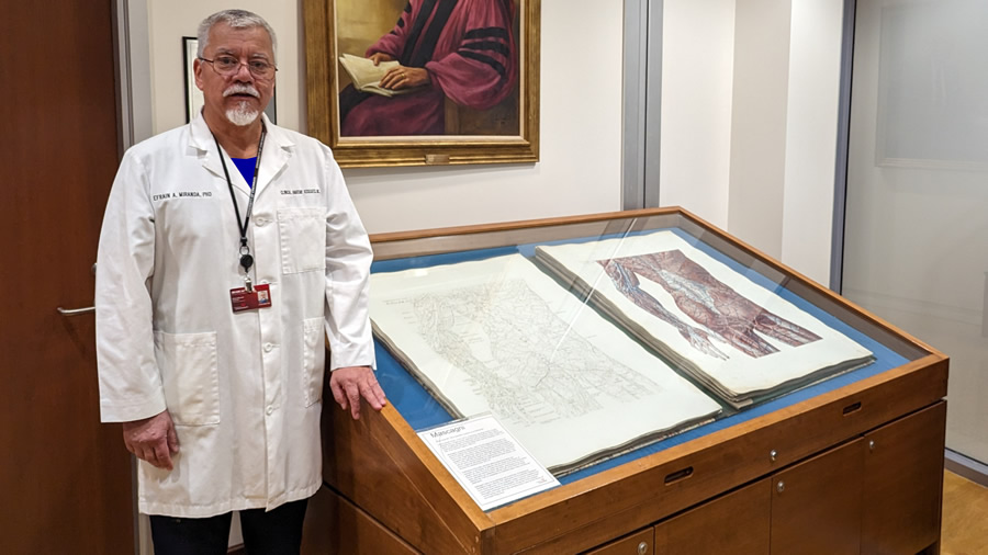
One of the individuals who piqued my interest was Henry Gray, FRS author of one of the great teaching books on anatomy ever written, “Gray’s Anatomy”. This book was first published in England in 1858, later published in the USA from 1862 to 1990. The English Edition is still published and is now in its 42nd Edition.

I started to look deeper into his life which, I learned with surprise, was not only very short, but obscure. Researching into Henry Gray’s life, I came about this book: “The Anatomist – A True Story of Gray’s Anatomy” by William (Bill) Hayes.

Bill Hayes started looking into Henry Gray’s life with the intent of writing a biography. He rapidly run into a wall. Besides a well-detailed list of academic titles and positions, degrees awarded, and scientific papers that he published. As the author states “what I had gathered about him would amount to little more than a Curriculum Vitae”. So, he starts a journey to uncover more data about Gray’s life and his well-known book.

Henry Gray, FRS (Public Domain)
Henry Gray, FRS

Henry Vandyke Carter, MD

Part of his journey was to understand the importance of anatomy as a component of the medical curriculum. Bill Hayes was accepted as a participant in the anatomy laboratory for physical therapy students and later visited medical students in the anatomy lab. Just his observations and comments throughout the book on these activities is worth reading this book.

What is interesting is the lack of personal information on Henry Gray. To this date, we do not have information about his birth, either location or date. There is more information on his family that on the subject. Most agree that Henry Gray was born in 1827, but some propose 1825. Either way, there is no firm data. What we do know is that he died on June 12, 1861, having contracted smallpox most probably from his nephew Charles Gray. Henry Gray was 36 years old.

Henry Gray and anatomy students
Gray and students

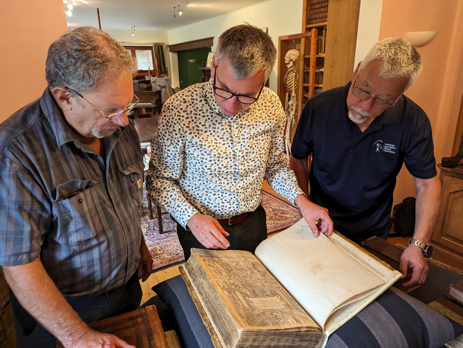
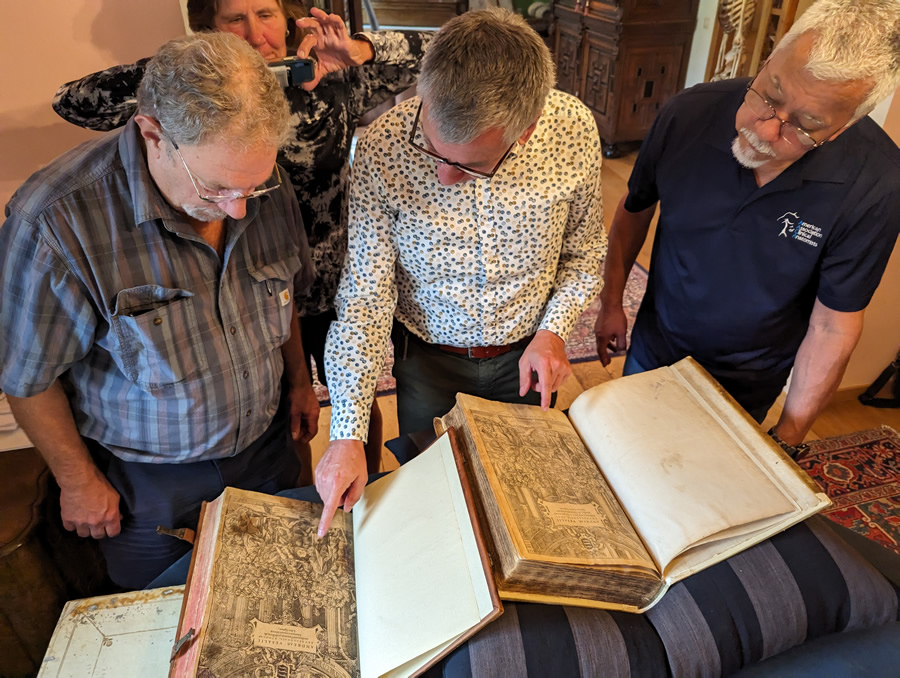
Here is one of the few photographs that exists of Henry Gray. In this particular 1870 picture by Joseph Langhorn, Gray is in the anatomy laboratory (forefront, third from the left) at St. George’s Hospital with his students. At the time it was customary for medical students to pose in the lab with bones and cadavers. This is a practice not in use today and disappeared circa 1930. For anyone interested in this now considered gruesome custom, I would recommend the book "Dissection" by John H. Warner and James M. Edmonson.

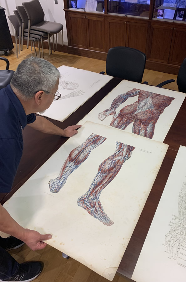
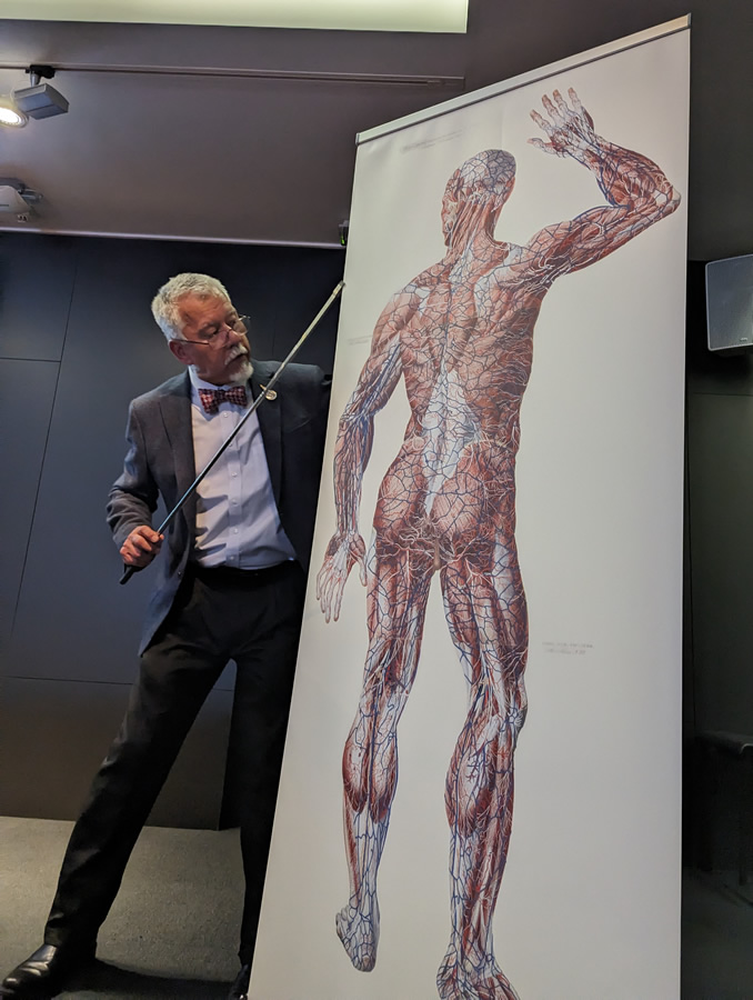
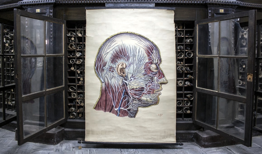
In his research, the author discovers Henry Gray’s collaborator and illustrator of the book: Henry Vandyke Carter, MD. Where Gray is obscure and with no personal information, Henry Carter writes a diary daily, and for some time he actually writes another diary that he calls “Reflections” on more personal and religious topics. Carter is a troubled, complicated individual blessed with incredible anatomical knowledge and drawing capabilities that can be seen throughout the book.

It is because of these diaries that we know Henry Carter and we can glimpse (almost at a distance) the character of Henry Gray, but it is not enough to elucidate his biography. In some ways it is like looking at an individual through a veil. You see him, but it is nebulous.

Much of the book is concentrated on Henry Carter, his life, his depression bouts, his self doubts and the work that he did illustrating Gray’s book. In some ways he is behind the scenes, and even though he did much of the dissection work and illustration, Henry Carter is mostly unknown to the anatomical world, as in may cases the medical illustrators are, with some notable exceptions.

Henry Carter is paid only 150 pounds for his work and even before getting paid he pays for a ticket to India where he accomplishes his objectives in life and in academia. Henry Carter eventually retires and comes back to England where he dies in 1897.

Bill Hayes and his partner visited rare book libraries at different universities and eventually go to London to visit areas and locations where Henry Gray lived and worked. There is a sense of accomplishment, but also a sense that we can almost touch the life of Henry Gray but fall short of seeing him.

The book ending is poignant, dealing with personal matters and thoughts on life and death. Eventually the author is very clear that the study of anatomy, although on dead subjects who donated their bodies to the universities is actually an activity that helps us understand life.

Bill Hayes is an awarded author of seven books, a frequent contributor to the New York Times and a photographer. More information about him and his activities at https://www.billhayes.com

Personal note: This is a book that I personally recommend and proud to add to my personal library. My one observation is that the author should have probably name the book “The Anatomists - A True Story of Gray’s Anatomy” as it is the story of the life of the two Henrys: Gray and Carter. In fact, I believe that Gray’s Anatomy could have been called “Gray’s and Carter’s Anatomy”, had history been slightly different – Dr. Miranda.

Sources:
1. “Henry Gray and Henry Vandyke Carter: Creators of a Famous Textbook" Roberts, S. J Med Biog 2000 8: 206-212
2. "Henry Gray, Anatomist: An Appreciation" Boland, F Am J Med Sci 1908 1827-1924
3. "The Anatomist: A True Story of Gray’s Anatomy" Hayes, B. Random House PG 2007